Our Services
The Breast Health Clinic makes your experience as pleasant and easy as possible. Our team is committed to providing you care in a supportive and healing environment.
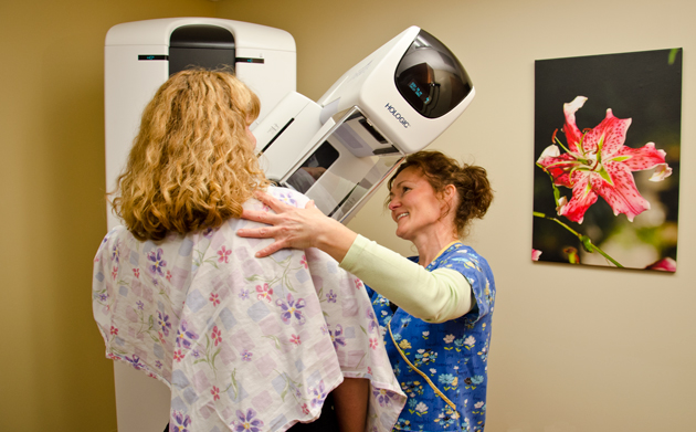
Digital Mammography
Mammography remains the single most effective tool in the detection of breast cancer. Studies have proven that the mortality rate from breast cancer is significantly reduced with regular screening mammograms.
3-D mammography (digital breast tomosynthesis): An advanced imaging technology that allows doctors to examine your breast tissue one layer at a time. 3-D mammography uses high-powered computing to convert digital breast images into a stack of very thin layers or “slices” – building what is essentially a “3-dimensional mammogram.”
A good analogy for 3-D mammography is like thinking of the pages in a book. If you look down at the cover you cannot see all the pages – but when you open it up, you can go through the entire book, page-by-page to see everything between the covers. 3-D mammography is designed with the same concept in mind.
Very low X-ray energy is used during the screening examination so your radiation exposure is below the FDA guidelines. Using 3-D mammography and digital mammography together for screening has been proven to significantly reduce “call-backs” by 20-40%. In addition, 3-D mammography finds cancers earlier than 2-D mammography alone with a 27% increase in cancer detection and a 40% increase in invasive cancer detection.
Because 3-D mammography offers earlier detection, Hillsboro Medical Center uses 3-D mammography for all patients as its primary screening method.
Ultrasound
A way to determine whether the mass seen on a mammogram or felt is solid versus cystic (malignant vs. benign). Ultrasound imaging uses high-frequency sound waves to generate images of the area of interest in your body.
Magnetic Resonance Imaging (MRI)
An imaging test that uses powerful magnets, radio waves and computer technology to create detailed images of the breast. It is capable of finding tiny tumors, even in dense breasts. MRI is sometimes used to look for breast cancer in women who are known to be at high risk. It can be used to stage cancers, map the extent of the tumor, evaluate the effectiveness of chemotherapy, and distinguish postoperative or post radiation scarring from recurrent cancer. MRI can also be used to image implants for leakage or ruptures.
Image-guided Biopsies
Even though imaging tests like the mammogram and ultrasound can find a suspicious area, they cannot tell whether it’s cancer. A biopsy is the only way to know for sure if a breast change is cancer. Lesions must be biopsied in the modality in which they are visualized; therefore we offer Stereotactic (mammography), ultrasound image-guided biopsies.
Bone Density Exams
Compared to many tests, the bone density exam is a breeze. It’s a touchless scan, similar to an X-ray, so it’s painless. You’ll wear clothing that contains no metal (zippers, metal buttons, and belts) around the waist area. The bone density test measures your hip, spine and/or wrist.
These tests are considered the “gold standard” for measuring your bone health. The bone density test is a highly sensitive scan and is more comprehensive than the peripheral machines so the results are more accurate. This makes the bone density scan the best way to tell if you have thinning bones. The test is easy to take; you lie down and the machine scans over your body in just a few minutes.